Research

Welcome to the Hummon Research Group at the Ohio State University. Our research interests lie at the intersection of analytical chemistry and chemical biology, with a focus on cancer biology. 


Current Projects:
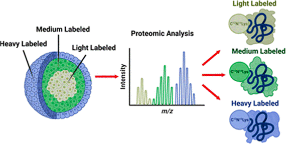
Spatial SILAC in Cellular Layers of the Spheroid

Stable isotopic labeling of amino acids in cell culture (SILAC) is a metabolic labeling method that identifies the origin of a protein in multiplexed experiments. By pulsing different heavy amino acids into cell culture media at set time points during spheroid growth, we can discretely label the cellular layers that form in these tumor models. Proteins from the necrotic core, quiescent middle layer, and proliferative outer layer of spheroids can be distinguished and quantified in a proteomic analysis, generating valuable information about chemotherapeutics’ effect on different cellular populations.
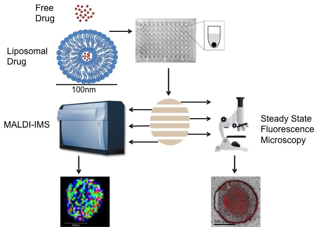
Analysis of Liposomal Drug Delivery Systems
Cancer chemotherapeutics often fail to reach all diseased cells.  To help solve this problem, researchers are investigating novel drug delivery systems.  Liposomes are an attractive option due to their low toxicity, high biocompatibility, and potential to carry a large amount of a drug to the tumor site, all while avoiding being eliminated from the body.  Using MALDI-IMS and fluorescence microscopy we analyze the spatial distribution of liposomal drugs in comparison to their free drug counterpart within spheroids. 

Investigation of Novel in vitro Platforms to Analyze the Efficacy and Toxicity of Chemotherapeutics
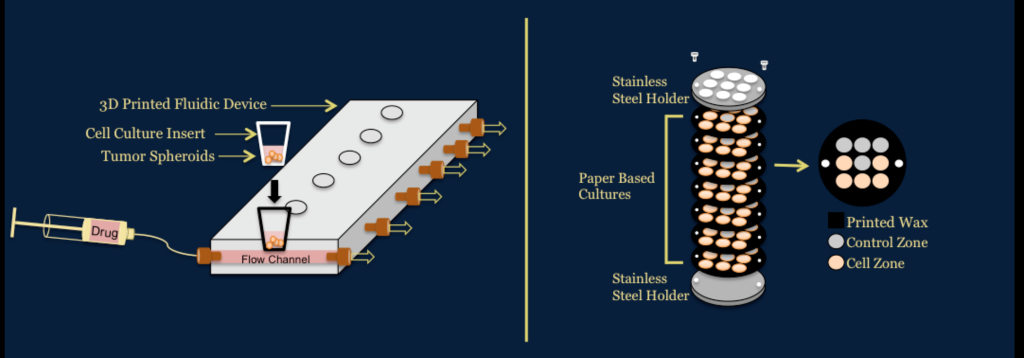
We use a number of platforms to determine the efficacy and toxicity of chemotherapeutics for colorectal cancer. One platform utilizes a 3D printed fluidic device developed in the Dr. Dana Spence laboratory at Michigan State University to dynamically dose spheroids.  The other platform utilizes paper based cultures developed in the Dr. Matthew Lockett laboratory at University of North Carolina at Chapel Hill.  These systems allow for applications to preclinical trials area of drug development pipeline. 

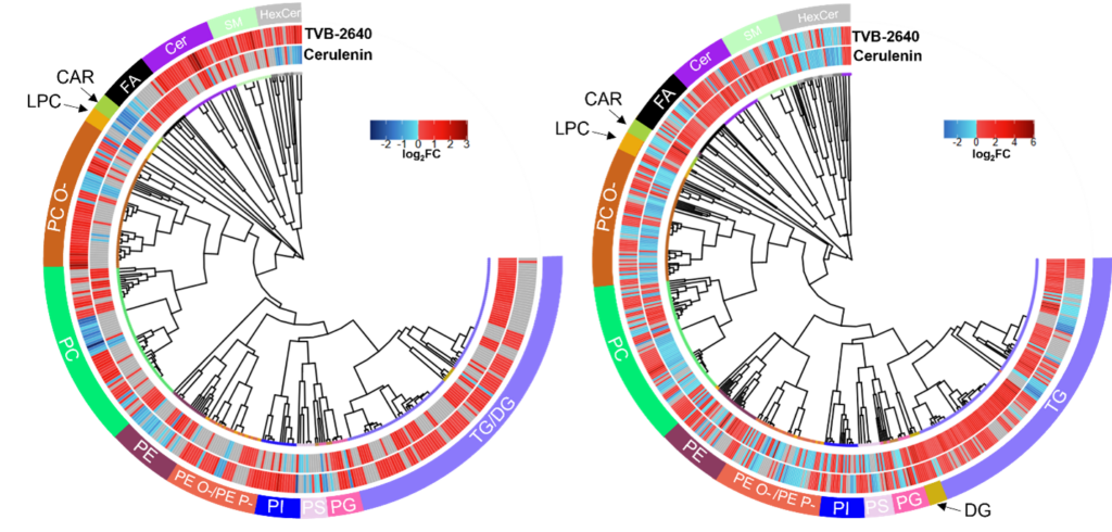
Lipidomics and proteomics of FASN inhibited colorectal cancer spheroids

FASN is often upregulated in a multitude of cancers and has become a druggable target of interest for the treatment of a multitude of cancers. However, the distinct molecular underpinnings of inhibiting this and other metabolic enzymes are unknown. Using both lipidomics and proteomics, we are able to determine the distinct molecular differences of different FAS inhibitors in two different sets of colorectal cancer spheroids and determine that each FAS inhibitor may be causing distinct death mechanisms to occur.
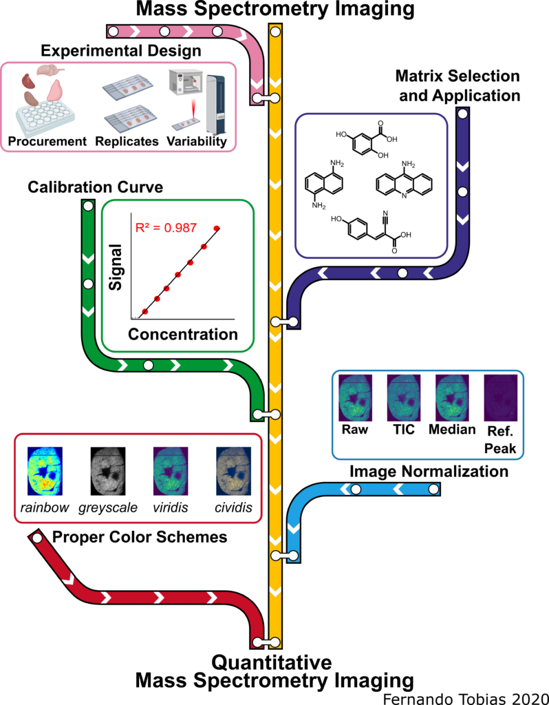
qMSI Scheme figure

Biomolecule Visualization and Quantification Using Mass Spectrometry Imaging 

The pink line represents experimental design and sources of variability, the purple line signifies the matrix selection process and application methods, the green line represents creating calibration curves for qMSI, the blue line symbolizes the image normalization aspect, while the red line represents the selection process for the proper color scheme of imaging data. The MS images depicted in this scheme are from the same murine heart section in a different color scheme and when the signal was normalized by different methods.